

Late nights, tiny organisms & questionable life choices
Conducting research at sea provides a unique opportunity to observe the plankton species you have known for years, alive and moving in seawater, freshly collected from their natural habitat and brought to the surface. Their natural colours and movements remain, something not to take for granted, especially when usually working with preserved samples.
In between laboratory work and filtering, we have found some extra time to seclude ourselves in the microscope room and take a look at samples that Anna has collected after her filtration work. When a long day’s work is done, our nights have settled into hot chocolate with Campurrianas’ cookies in the company of late-night colleagues, followed by some time getting better at ping pong and observing our samples under the microscope (yes, just for fun).

Most of the organisms we observe are phytoplankton. These single-celled organisms might be small, but remarkably important, contributing to over half of the global primary production. Many of them are auto- or mixotrophs, meaning that they either rely exclusively on photosynthesis for their energy supply or combine photosynthesis with other ways of obtaining it. One group widely considered to be primarily autotroph are diatoms– our favorite. Diatoms are beautiful, symmetrical organisms that can occur in solitary cells, chains and colonies. Another group regularly found in phytoplankton samples are dinoflagellates, which are often heterotrophic or mixotrophic and come in a fascinating variety of shapes. With their two flagella, they can swim to a certain degree, which can be entertaining to watch under the microscope. We’ve seen dinoflagellates belonging to the family Gymnodinaceae moving by constantly rotating their bodies.
During the expedition, we’ve already encountered many species that we had only seen in books before. For example, we found a few cells of Coscinodiscus cf. radiatus (Fig. 2C) and Asteromphalus (Fig. 2K), many dinoflagellates of the genus Tripos (Fig. 2J, M) and various tintinnids (Fig. 2D). However, occasionally, we also find organisms whose identity remains unknown. For instance, we found a mysterious star shaped cell that could allegedly be an internal structure of the dinoflagellate Actiniscus pentasterias, which becomes visible once the cell degrades. This star made of biogenic silica can become part of the microfossil record (Fig. 2H).
Every station we sample is different and some surprise us with a diversity of diatoms that make the perfect model for a photoshoot. Even if we have mastered the art of taking photos through the objective with the phone, taking them while the ship is moving has proved to be a little more challenging, since the cells often seem to be having a rollercoaster ride.

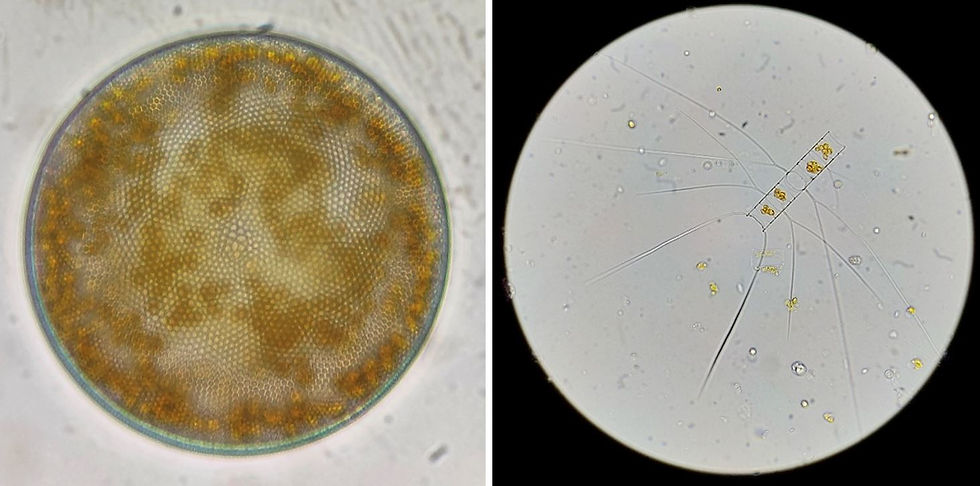

As the base of marine food webs, phytoplankton are an important food source for small zooplankton, most notably copepods. On board our research vessel, we also have a zooplankton expert, Marja, who every now and then sets aside zooplankton samples for us to observe, and has explained how to tell apart the most common copepod species found in these waters (Fig 5). We have enjoyed watching the bluish spark of bioluminescent copepod Metridia and the hypnotic dance of pteropods that Catarina enjoys so much. Since zooplankton are usually bigger than phytoplankton, we use a binocular instead of a microscope. They are huge in this area! Visible to the naked eye, some as big as a fingernail. Sometimes though, even the binocular has high enough magnification to distinguish larger diatoms or diatom chains. The centric diatom genus Coscinodiscus, for example, includes species that can reach 500µm (=0.5mm).


Comments